
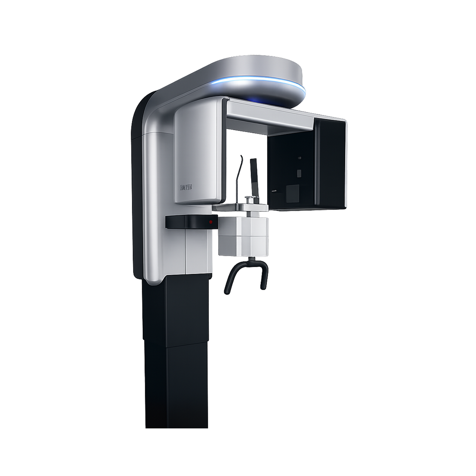
PreXion 3D Explorer EX(Old Version)
The powerful system components of the PreXion 3D Explorer enable an extraordinary combination of the most precise 3D imaging, large image detail, lowest radiation exposure, reliable diagnostics, and digital planning for all indications in modern dentistry.
Choose options
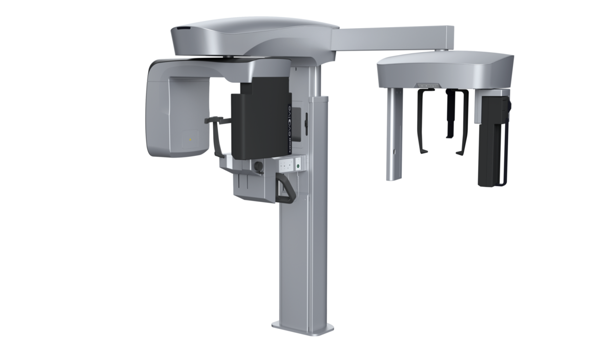
PreXion 3D Explorer EX
The powerful system components of the PreXion3D Explorer enable an extraordinary combination of the most precise 3D imaging, large image detail, lowest radiation exposure, reliable diagnostics and digital planning for all indications in modern dentistry, including periodontology, endodontics, implantology, orthodontics and maxillofacial surgery, among many others. Its patient management system is designed for secure and networked communication of patient data across multiple rooms within a practice and can be integrated into the existing infrastructure with ease.
With the precision and professional competence of PreXion machine, dental professionals have a powerful partner at their side.
Software Features
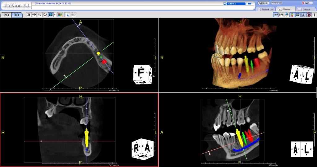
• Multi-data – Load multiple patient scans on a single screen. Synchronize pre- and post-operative scans and detect differences, slice-by-slice.
• Patient Education and Presentation – Quickly capture 3D animated video clips for patient education, case acceptance and lecture presentations. Increase case acceptance through better patient understanding.
• Collaborative Tools – Automatically save 3D image reports to MS Word template and attach to patient’s practice management record. Collaborate with referring dentists by burning a patient disc with sample viewer. Capture and email images quickly.
• Remote Access – Work on cases from home or a satellite office without long connectivity delays. Lead virtual online treatment planning meetings remotely with PreXion Cone Beam.
• Thin Client Server – PreXion3D CBCT scanners do not require computer hardware upgrades. It does not slow down network bandwidth like other CBCT scanners.
• Implant Library – Use our extensive library or customize your own library.
• Save Scenes – Save your case workup as a scene or create multiple saved scenes with a single scan.
• 3D Templates – Save time with over 20 pre-made 3D volume rendering templates or customize your own.
• Slab and Cutting – Slab feature allows the clinician to see inside structures while rotating the 3D image. Cutaway structures to see exactly what is pertinent to your study.
See it in Action
Clinical Applications
- Implant placement surgery
- Endodontics
- Periodontics
- Oral-maxillofacial surgery
- TMJ
- Pathology
- Impacted and supernumerary teeth
- DICOM export for implant surgical guides & CAD/CAM integration
- Airway analysis
Highlights
- Accurate 360-degree gantry rotation
- 260-1,024 projected views
- Dedicated 2D pan mode option
- Clearest detail with 0.3mm focal spot & 0.08-0.2mm voxel
Product Comparison
| Device Type | Cone Beam Computed Tomography System, Head/Neck |
| Focal Spot | 0.3 mm x 0.3 mm |
| Tube Voltage | 90–110 kV |
| Tube Current | 1–5 mA |
| Total Filtration | 2.7 mm AI |
| Voxel Size | 0.07 mm, 0.1 mm, 0.2 mm, 0.3 mm |
| FOV | Teeth: 5×5 cm Arch: 10×10 cm Full: 15.8 cm Face: 15x10cm |
| Standard Mode | 10 seconds |
| High Definition Mode (HD) | 18 seconds |
| Ultra High Definition Mode (UHD) | 20 seconds |
| Panoramic Standard Mode | 14 seconds |


All Practice Solutions is a trusted provider of high-quality dental solutions. We specialize in CBCT scanners, dental microscopes, digital imaging solutions, dental chairs, X-ray generators, and more to meet the needs of modern dental practices. Additionally, we offer expert digital marketing services, making us a top SEO company and website design partner for dentists nationwide. Contact us today for innovative solutions and expert services!
Powered by
 Sitemap © Copyright 2026 - All Rights Reserved
Privacy Policy |
Terms and Conditions
Sitemap © Copyright 2026 - All Rights Reserved
Privacy Policy |
Terms and Conditions
