
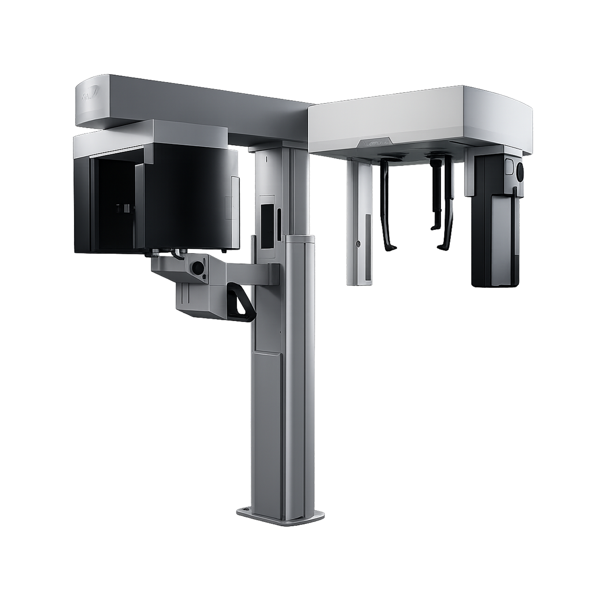
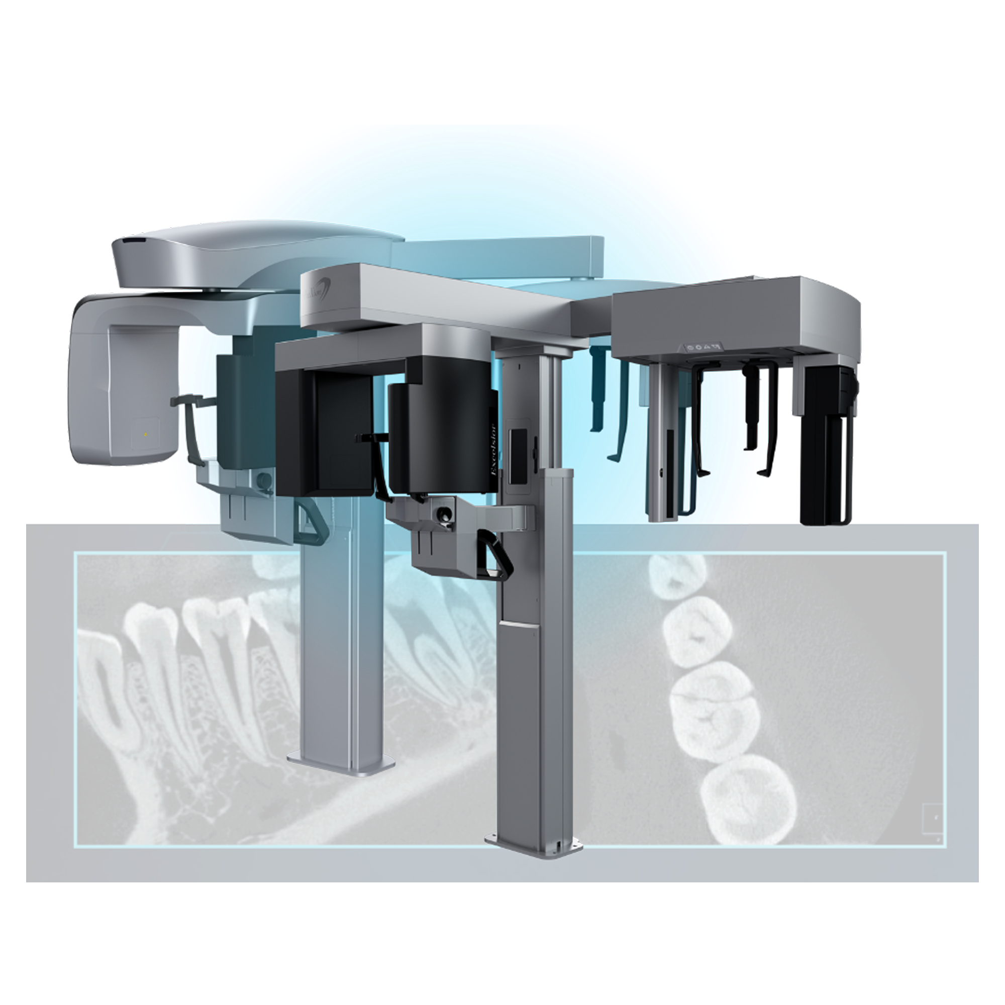
PreXion Excelsior MID (MFOV)
The Evolution of 3D Dental Imaging in a Technology Product
A versatile mid-field CBCT system built for comprehensive diagnostics across multiple dental specialties. It delivers detailed 3D imaging with low radiation exposure and strong data management capabilities.
Choose options
Update Your Technology in 2026 with the PreXion Excelsior Mid
The PreXion Excelsior Mid scanner is the evolution of our original flagship product, designed for dentists who demand the highest diagnostic clarity and detail.
Why choose the PreXion Excelsior Mid?
The powerful system components of the PreXion Excelsior Mid enable an extraordinary combination of the most precise 3D imaging, high image detail, lowest radiation exposure, reliable diagnostics, and digital planning for all indications in modern dentistry, including periodontology, endodontics, implantology, orthodontics, maxillofacial surgery, and more. Its patient management system is designed for secure and networked communication of patient data across multiple rooms within a practice and can be integrated into the existing infrastructure with ease.
Software Features
With the precision and professional competence of PreXion, dental professionals have a powerful partner at their side.
2D Reports
Our 2D Reports feature for panoramic and cephalometric images allows customizable annotations with arrows, circles, and color highlights. It also enables precise implant planning directly on the panoramic view, improving clarity and documentation.
2D Manual and AI-Assisted Cephalometry
Our 2D Cephalometry feature allows clinicians to perform cephalometric tracing either manually or with automated landmark detection powered by AI. This accelerates workflows, improves accuracy, and ensures consistent measurements for orthodontic evaluation and treatment planning.
3D Implant Planning
Our implant planning module allows users to mark the mandibular canal and position implant fixtures from an extensive library of real-size systems. Based on the simulation, clinicians can determine ideal implant dimensions, depth, and angulation. Compatible workflows also support the ordering of highly accurate surgical guides.
Airway Volume Measure
The segmentation tool allows users to obtain volumetric data by isolating structures based on density values. It can be used to segment airways, sinuses, the mandible, and teeth, automatically providing corresponding volume measurements.
STL Conversion
Convert DICOM data into STL files for seamless use in CAD/CAM workflows and 3D printing. This tool enables accurate model generation for planning, simulations, and additive manufacturing.
Virtual Endoscopy
By combining CPR (Curved Planar Reformat) with virtual view technology, the software enables virtual endoscopy. Users simply define a navigation path, and the system generates an internal airway view for detailed examination.
DICOM Fusion
The Fusion tool allows users to align and overlay multiple DICOMs for more accurate diagnostics and planning. This unified view enhances anatomical correlation and supports precise clinical decision-making.
3D Reports
The reporting tool offers flexible templates that simplify clinical documentation, allowing users to create custom layouts and drag-and-drop images with ease. Reports can be saved locally or in the database in formats such as HTML, PPT, or PDF.

16 Years of
Experience

Clearest
Images

Clinical
Advisory Board

Distributing
Partners

Customer
Support
Field of View


3D Algorithms: Enhancing Your Workflow Efficiency and Diagnostic Accuracy
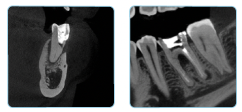
UHD Mode for Endodontics
PreXion Excelsior MID has an isotropic voxel resolution of 75 μm to 250 μm.
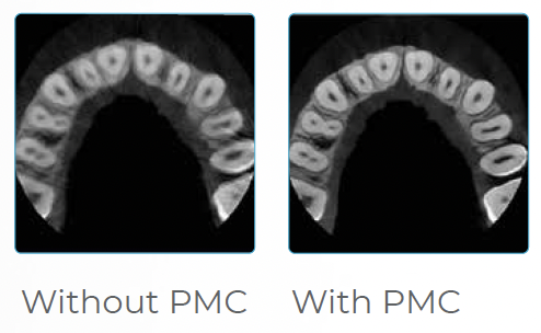
Patient Motion Correction (PMC)
The PreXion Evolve algorithm automatically corrects the image imperfections caused by patient micro-movements. This ensures high exam quality, avoids repetition, and offers greater accuracy in making diagnoses.
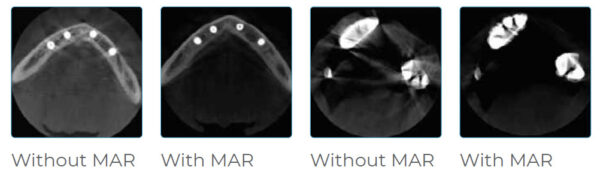
Metal Artifact Reduction (MAR)
The PreXion Evolve algorithm automatically corrects the image imperfections caused by patient micro-movements. This ensures high exam quality, avoids repetition, and offers greater accuracy in making diagnoses.


All Practice Solutions is a trusted provider of high-quality dental solutions. We specialize in CBCT scanners, dental microscopes, digital imaging solutions, dental chairs, X-ray generators, and more to meet the needs of modern dental practices. Additionally, we offer expert digital marketing services, making us a top SEO company and website design partner for dentists nationwide. Contact us today for innovative solutions and expert services!
Powered by
 Sitemap © Copyright 2026 - All Rights Reserved
Privacy Policy |
Terms and Conditions
Sitemap © Copyright 2026 - All Rights Reserved
Privacy Policy |
Terms and Conditions
